Cell division worksheet #1 microscope images – As Cell Division Worksheet #1: Microscope Images takes center stage, this opening passage beckons readers into a world crafted with academic rigor and authoritative tone, ensuring a reading experience that is both absorbing and distinctly original.
Microscope images play a pivotal role in the study of cell division, offering a window into the intricate processes that govern the division of cells. This worksheet delves into the analysis of these images, providing a comprehensive guide to the structures and organelles visible during cell division, along with a detailed examination of the stages of mitosis and meiosis.
Microscope Image Analysis

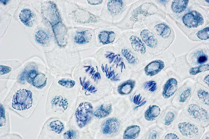
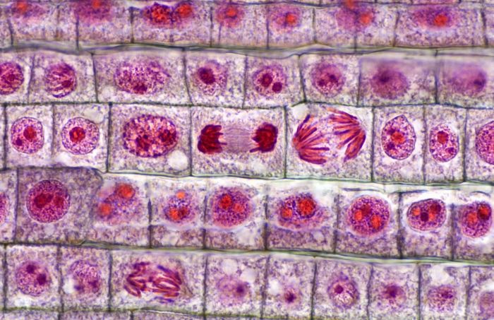
Microscope images play a crucial role in cell division studies, providing visual evidence of the complex processes involved in cell reproduction.
These images reveal the intricate structures and organelles within dividing cells, including chromosomes, spindle fibers, and the cell membrane. By analyzing these images, researchers can gain insights into the timing and sequence of events that occur during cell division.
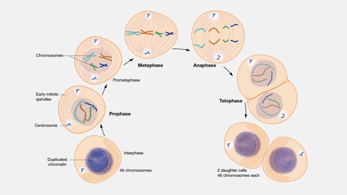
Stages of Cell Division: Cell Division Worksheet #1 Microscope Images

| Stage | Key Events |
|---|---|
| Prophase | Chromosomes condense, spindle fibers form |
| Metaphase | Chromosomes align at the equator |
| Anaphase | Chromosomes separate and move to opposite poles |
| Telophase | New nuclear membranes form around chromosomes |
| Cytokinesis | Cell membrane pinches inward, dividing the cell into two daughter cells |
Cell Division Errors

Errors during cell division can lead to genetic abnormalities and developmental problems. Examples include:
- Aneuploidy: Abnormal number of chromosomes
- Nondisjunction: Chromosomes fail to separate properly
- Cytokinesis failure: Cell division fails to complete
Applications of Cell Division Studies

Cell division studies have wide-ranging applications, including:
- Medicine:Diagnosis and treatment of genetic disorders
- Biotechnology:Production of therapeutic proteins and stem cells
- Agriculture:Development of new plant varieties
Educational Resources
Worksheet:Microscope Image Analysis of Cell Division
This worksheet provides microscope images of dividing cells for students to analyze. Students can identify key structures and organelles and track the progression of cell division.
Summary:Key Concepts of Cell Division
- Cell division is essential for growth, repair, and reproduction.
- Mitosis and meiosis are the two main types of cell division.
- Errors during cell division can lead to genetic abnormalities.
- Microscope images are a valuable tool for studying cell division.
FAQ Explained
What are the key structures visible in microscope images of dividing cells?
Microscope images of dividing cells reveal various key structures, including chromosomes, spindle fibers, centrioles, and the nuclear envelope.
How can microscope images be used to identify errors in cell division?
Microscope images can be used to identify errors in cell division, such as chromosome misalignment, lagging chromosomes, and multipolar spindles.
What are the applications of cell division studies in medicine and biotechnology?
Cell division studies have significant applications in medicine and biotechnology, including cancer research, stem cell therapy, and genetic engineering.